

UT analytical chemists, microbiologists and engineers are collaborating to use 3-D printing technology to create protein houses for bacteria. Researchers use this method to better determine how different environmental changes affect
the bacteria.
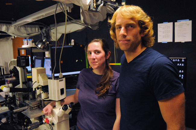
Postdoctoral fellow Jodi Connell is the primary researcher using these methods to study which bacteria can develop
antibiotic resistance.
Connell said studying bacteria via traditional research methods — shaking them in a flask, for example — can help researchers study different genes. Using the 3-D cages, researchers are able to isolate the bacteria and control their environment and inter-species interactions. This allows Connell to then study these interactions within the controlled environment and specifically how they
cause infections.
“What students probably do not realize is that bacteria are actually very social creatures,” molecular biosciences professor Marvin Whiteley said.
Connell said bacteria can gauge the number and type of other bacteria around them by secreting and sensing chemical signals. Bacteria can communicate with each other by coordinating gene expression to display a set of characteristics when they are around other bacteria that they may not when they are isolated.
“No one really knows the spatial requirements … or what induces these behaviors physically,” Connell said.
Postdoctoral researcher Eric Ritschdorff described the basic process of using the 3-D cages to study bacteria. Researchers mix gelatin with other molecules and cells, use a laser focus to define an orientation and then wash away the gelatin that is not reactive to light. This leaves cells inside a container that resarchers can then study, Ritschdorff said.
Whiteley said Connell’s major challenge was to make the systems biocompatible with, or safe for, the bacteria.
Ritschdorff said another difference between this and previous research done using the method is earlier research relied on creating a house before adding cells.
“The benefit of this method is [Connell] can define the orientation or placement of bacteria any way she wants,” Ritschdorff said.
Whiteley said this method can be applied to researching antibiotic resistance, which scientists are only beginning to understand.
“No one really knows how bacterial infection actually works,” Whiteley said.
When researchers study bacteria in a test-tube, there are limits to what they can learn because bacteria encounter certain kinds of stress in this environment. For example, bacteria in test tubes run out of food and accumulate waste products, limiting their density. Bacteria in nature exist in little communities, and using 3-D cages allows researchers to roughly simulate these communities and study how they may be a major mode of disease transmission.
Whiteley said within the next few years he and other researchers hope to answer these questions and use this method in animals to create models of infection
and disease.
“We are learning now that you may not need millions of cells to feed an infection,” Connell said. “It may be 50 [cells that] become resistant to the antibiotics.”